NCERT Solutions for Class 11 Biology Chapter 6 (2025-2026)
Chapter 6 of Class 11 Biology -Anatomy of Flowering Plants -takes students beneath the surface of plants to explore their internal structure and organization. This chapter explains how different plant tissues like meristematic and permanent tissues function together to support growth, transport, and survival. It also covers the anatomy of roots, stems, and leaves in both dicot and monocot plants, highlighting the differences and similarities that help plants adapt to their environment.This chapter is part of the comprehensive NCERT Solutions Class 11 Biology series, which covers all chapters in detail.
The NCERT Solutions for Anatomy of Flowering Plants provided here include clear, step-by-step answers to all textbook questions. These solutions help students grasp complex diagrams, understand structural details, and build a strong foundation for exams — especially essential for NEET and other competitive tests that emphasize plant anatomy concepts.
NCERT Solutions for Class 11 Biology Chapter 6 - Questions & Answers
Class 11 Chapter 6 Biology Questions & Answers –Anatomy of Flowering Plants
Q1. State the location and function of different types of meristems.?
Solution: Depending on the location in plants, meristems are classified into the following three categories:
| S.No. | Type of meristem | Location and Function |
| 1. | Shoot apical meristem | It is present in the apical tissue of shoot and is responsible for the vertical growth and elongation of a plant. |
| 2. | Root apical meristem | It is present in the tip region of root and is responsible for root growth. |
| 3. | Intercalary meristem | It is present in between the permanent tissues and is observed in grasses. Its main function is to regenerate the damaged plant part. |
|
4.
|
Lateral meristem
|
It is present in the mature regions of shoots and roots. It is responsible for the production of secondary tissues.
|
Q2. Cork cambium forms tissues that form the cork. Do you agree with this statement? Explain.?
Solution: During the secondary growth, the plant increases its girth as a result of the activity of vascular cambium. This leads to the rupturing of the outer cortical and epidermal layer. To protect the internal tissues from the exposure to the external environment, another meristematic tissue known as cork cambium develops in the cortex region. This tissue is a couple of layers thick and is made up of rectangular thin-walled cell. The outer cells divide to form cork and the cells of the inner layer form secondary cork cambium. Thus, it can be said that the cork cambium forms tissues that give rise to the cork.
Q3. Explain the process of secondary growth in the stems of woody angiosperms with the help of schematic diagrams. What is its significance?
Solution: In the stems of woody angiosperms, a meristematic layer called intrafascicular cambium is present between the primary xylem and phloem. During the growing season, the cells of medullary rays adjoining the intrafascicular cambium become meristematic and form a continuous ring that divides bidirectionally. The cell that cut off toward the pith becomes secondary xylem, while the cell that cut outwardly form secondary phloem. The activity on the cambial ring is high inwardly, resulting in the production of more secondary xylem, compared to secondary phloem. This results in the formation of a compact mass at the centre of the stem.
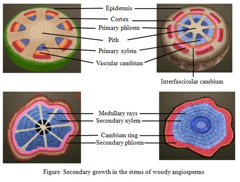
Significance: Many physiological and environmental factors govern the activity of intrafascicular cambium. This can be distinctly observed in woody trees of temperate regions. They have clearly demarcated annual rings which are concentric rings formed due to the formation of two different kinds of woods during two different seasons. In the spring, cambium is active and produces a large number of xylary elements having vessels with wider cavities. This wood is called springwood and is lighter in colour with lower density. During the winter season, cambium is less active and forms fewer xylary elements that have narrow vessels. This latewood or autumn wood is darker in colour and has a higher density. This difference in the woods formed during two different seasons results in the formation of annual rings. One can find out the age of the plant, based on the number of annual rings seen in the cut stem of the plant.
Q4. Draw illustrations to bring out the anatomical difference between
(a) Monocot root and Dicot root
(b) Monocot stem and Dicot stem
|
Monocot root
|
Dicot root
|
|
The roots grow randomly into the ground.
|
The roots grow straight down and then branch out.
|
|
The central pith is large and very well developed.
|
Pith is comparatively small.
|
|
Does not show secondary growth
|
Shows secondary growth
|
|
More than six xylem bundles are seen.
|
Few xylem bundles are seen in dicot root (less than six).
|
 |
 |
|
Monocot stem
|
Dicot stem
|
|
Hypodermis consists of sclerenchymatous cells.
|
Hypodermis consists of few layers of collenchymatous cells.
|
|
Vascular bundles are scattered in ground tissue.
|
Vascular bundles are arranged in a ring.
|
|
Vascular bundles are closed.
|
Vascular bundles are open.
|
|
Starch sheath, pericycle, pith and medullary rays are absent.
|
Starch sheath, pericycle, pith and medullary rays are present.
|
|
Phloem parenchyma is absent.
|
Phloem parenchyma is present.
|
 |
 |
Q5. Cut a transverse section of young stem of a plant from your school garden and observe it under the microscope. How would you ascertain whether it is a monocot stem or a dicot stem? Give reasons.
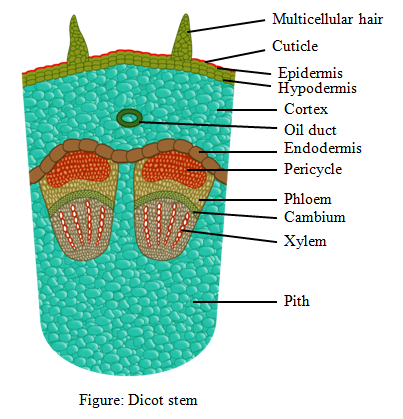
Solution: The well-differentiated ground tissue with the arranged vascular bundle is the characteristic feature of dicot stem while undifferentiated ground tissue and the scattered vascular bundle is the feature of monocot stem. If the cut section shows the presence of vascular bundle arranged in a ring, it is a dicot stem but if the vascular bundles are scattered, it is a monocot stem.
Q6. The transverse section of a plant material shows the following anatomical features - (a) the vascular bundles are conjoint, scattered and surrounded by a sclerenchymatous bundle sheath. (b) phloem parenchyma is absent. What will you identify it as?
Solution: Conjoint, scattered and closed vascular bundles surrounded by bundle sheath cells, are some of the characteristic anatomical features of a monocot stem. Medullary rays as well as phloem parenchyma are also absent in these stems.
Q7. Why are xylem and phloem called complex tissues?
Solution: The tissues which are composed of more than one type of cells working together to bring about a function are called complex tissues. Both xylem and phloem are made up of more than one type of cells, thus they are known as complex tissues.
The different types of elements present in the xylem tissue are:
- Tracheids,
- Vessels,
- Xylem fibres and
- Xylem parenchyma.
The different types of elements present in the phloem tissue are:
- Sieve tube elements,
- Companion cells,
- Phloem parenchyma and
- Phloem fibres
Q8. What is stomatal apparatus? Explain the structure of stomata with a labelled diagram.
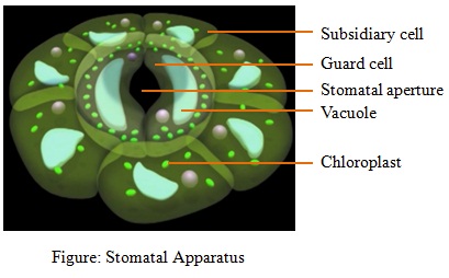
Solution: The stomatal aperture/pore and guard cells along with the subsidiary cells form the stomatal apparatus. Stomata are the special openings present in the epidermis of the leaves to facilitate gaseous exchange and transpiration. Stomata are made up of two kidney-shaped (bean-shaped) guard cells and the turgidity of these cells regulate the opening and closing of the stomatal pore. The cell wall of guard cells shows differential thickening. The outer wall is elastic and thin, while the inner wall that is close to the pore is very thick. These guard cells are photosynthetic in nature. The guard cells are surrounded by specialized epithelial cells called subsidiary cells which have a specific shape and size.
Q9. Name the three basic tissue systems in the flowering plants. Give the tissue names under each system.
Solution: The three basic tissue systems in the flowering plants are as follows:
| Tissue System | Tissue Names |
| Epidermal tissue system | Stomata, root hairs and trichomes and epidermis |
| Ground tissue system | Parenchyma, sclerenchyma, collenchyma and mesophyll |
| Vascular tissue system | Phloem, cambium and xylem |
Q10. How is the study of plant anatomy useful to us?
Solution: Study of plant anatomy is useful in the following ways:
- Classification: On the basis of anatomical features, a plant can be classified into monocot or dicot.
- Adaptation: Anatomy reveals about the various modifications in the internal organization of tissue with respect to different environmental conditions; thus, it helps in studying evolution.
- Economic value: Anatomical study suggests the quality of wood in the plant and helps in predicting the quality of fibres that can be obtained from the plants.
Q11. What is periderm? How does periderm formation take place in the dicot stems?
Solution: During secondary growth, the plant increases its girth due to the activity of vascular cambium. This results in rupturing of the outer cortical and epidermal layer. To protect the internal tissue from the external environmental exposure, the meristematic tissue develops in this region, called cork cambium or phellogen. This meristematic tissue possesses a rectangular thin-walled cell, which divides outwardly to form cork or phellem and inwardly to form secondary cork cambium phelloderm. This tissues, namely phellogen, phellem and phelloderm, are collectively called periderm. Periderm formation in dicot stem takes place from the meristematic tissue of the cortex region.
Q12. Describe the internal structure of a dorsiventral leaf with the help of labelled diagrams.
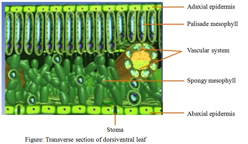
Solution: The dicot leaves differ in the structure and appearance of its upper and lower surfaces, thus they are called dorsiventral leaves. The vertical section of dorsiventral leaves under a microscope can be clearly distinguished into three parts. They are:
- Epidermis: This is the outermost layer of tissue that covers the upper and lower surfaces of the leaf. The epidermis at the upper surface is called adaxial epidermis while that at the lower surface is called abaxial epidermis. Generally, the lower epidermis contains more stomatal opening while upper epidermis contains very few or no stomata.
- Mesophyll: The mesophyll cells appear green as they contain chloroplast. This photosynthetic tissue is present between the upper and lower epidermis. The elongated mesophyll cells, arranged vertically in parallel direction are called palisade parenchyma. These are close to the upper epidermis. Below palisade parenchyma are loosely arranged oval cells, known as spongy parenchyma that extends down to lower epidermis.
- Vascular system: These include densely arranged complex tissues like xylem and phloem.
More Resources of NCERT Solutions for Class 11 Biology
- NCERT Solutions for Class 11 Biology Chapter 1
- NCERT Solutions for Class 11 Biology Chapter 2
- NCERT Solutions for Class 11 Biology Chapter 3
- NCERT Solutions for Class 11 Biology Chapter 4
- NCERT Solutions for Class 11 Biology Chapter 5
- NCERT Solutions for Class 11 Biology Chapter 7
- NCERT Solutions for Class 11 Biology Chapter 8
- NCERT Solutions for Class 11 Biology Chapter 9
- NCERT Solutions for Class 11 Biology Chapter 10
- NCERT Solutions for Class 11 Biology Chapter 11
- NCERT Solutions for Class 11 Biology Chapter 12
- NCERT Solutions for Class 11 Biology Chapter 13
- NCERT Solutions for Class 11 Biology Chapter 14
- NCERT Solutions for Class 11 Biology Chapter 15
- NCERT Solutions for Class 11 Biology Chapter 16
- NCERT Solutions for Class 11 Biology Chapter 17
- NCERT Solutions for Class 11 Biology Chapter 18
- NCERT Solutions for Class 11 Biology Chapter 19
NCERT Solutions for Class 11 Biology Chapter 6 - FAQs
1. What topics are covered in Chapter 6 of NCERT Class 11 Biology?
Chapter 6 introduces the internal structure and functional organisation of flowering plants. It covers the tissues, the tissue system, anatomical differences in dicot and monocot roots, stems and leaves, and secondary growth in plants.
2. How do the NCERT Solutions for Chapter 6 help students prepare for exams?
The solutions offer step-by-step answers, labelled diagrams, explanation of anatomical features and differences, and align with the textbook’s exercise questions. They help clarify concepts, practise diagrams, and perform well in school and competitive exams.
3. What are the three basic tissue systems in flowering plants according to this chapter?
The three basic tissue systems are:
- Epidermal tissue system
- Ground tissue system
- Vascular tissue system
4. Why is secondary growth discussed in Chapter 6, and what is its significance?
Secondary growth refers to the increase in girth or thickness of plant stems/roots, particularly in dicots, through the activity of lateral meristems (vascular cambium and cork cambium). It provides mechanical support, additional vascular tissue, and protection (via periderm) to the plant.
Q.1 State the location and function of different types of meristems.
Ans-
Depending on the location in plants, meristems are classified into the following three categories:
| S.No. | Type of meristem | Location and Function |
| 1. | Shoot apical meristem | It is present in the apical tissue of shoot and is responsible for the vertical growth and elongation of a plant. |
| 2. | Root apical meristem | It is present in the tip region of root and is responsible for root growth. |
| 3. | Intercalary meristem | It is present in between the permanent tissues and is observed in grasses. Its main function is to regenerate the damaged plant part. |
| 4. | Lateral meristem | It is present in the mature regions of shoots and roots. It is responsible for the production of secondary tissues. |
Q.2 Cork cambium forms tissues that form the cork. Do you agree with this statement? Explain.
Ans-
During the secondary growth, the plant increases its girth as a result of the activity of vascular cambium. This leads to the rupturing of the outer cortical and epidermal layer. To protect the internal tissues from the exposure to the external environment, another meristematic tissue known as cork cambium develops in the cortex region. This tissue is a couple of layers thick and is made up of rectangular thin-walled cell. The outer cells divide to form cork and the cells of the inner layer form secondary cork cambium. Thus, it can be said that the cork cambium forms tissues that give rise to the cork.
Q.3 Explain the process of secondary growth in the stems of woody angiosperms with the help of schematic diagrams. What is its significance?
Ans-
In the stems of woody angiosperms, a meristematic layer called intrafascicular cambium is present between the primary xylem and phloem. During the growing season, the cells of medullary rays adjoining the intrafascicular cambium become meristematic and form a continuous ring that divides bidirectionally. The cell that cut off toward the pith becomes secondary xylem, while the cell that cut outwardly form secondary phloem. The activity on the cambial ring is high inwardly, resulting in the production of more secondary xylem, compared to secondary phloem. This results in the formation of a compact mass at the centre of the stem.

Significance: Many physiological and environmental factors govern the activity of intrafascicular cambium. This can be distinctly observed in woody trees of temperate regions. They have clearly demarcated annual rings which are concentric rings formed due to the formation of two different kinds of woods during two different seasons. In the spring, cambium is active and produces a large number of xylary elements having vessels with wider cavities. This wood is called springwood and is lighter in colour with lower density. During the winter season, cambium is less active and forms fewer xylary elements that have narrow vessels. This latewood or autumn wood is darker in colour and has a higher density. This difference in the woods formed during two different seasons results in the formation of annual rings. One can find out the age of the plant, based on the number of annual rings seen in the cut stem of the plant.
Q.4 Draw illustrations to bring out the anatomical difference between
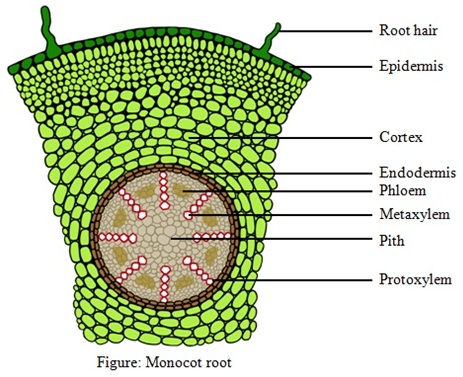
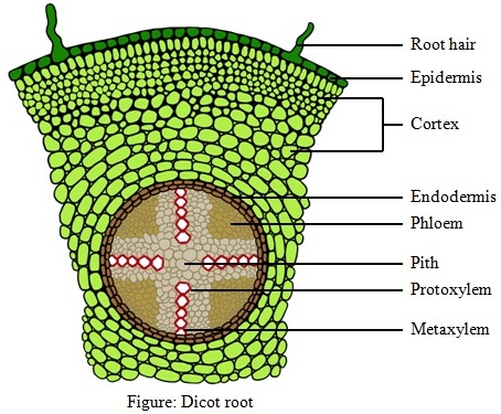
(a) Monocot root and Dicot root
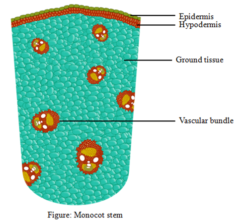
(b) Monocot stem and Dicot stem
Ans-
| Monocot root | Dicot root |
| The roots grow randomly into the ground. | The roots grow straight down and then branch out. |
| The central pith is large and very well developed. | Pith is comparatively small. |
| Does not show secondary growth | Shows secondary growth |
| More than six xylem bundles are seen. | Few xylem bundles are seen in dicot root (less than six). |
 |
 |
| Monocot stem | Dicot stem |
| Hypodermis consists of sclerenchymatous cells. | Hypodermis consists of few layers of collenchymatous cells. |
| Vascular bundles are scattered in ground tissue. | Vascular bundles are arranged in a ring. |
| Vascular bundles are closed. | Vascular bundles are open. |
| Starch sheath, pericycle, pith and medullary rays are absent. | Starch sheath, pericycle, pith and medullary rays are present. |
| Phloem parenchyma is absent. | Phloem parenchyma is present. |
 |
 |
Q.5 Cut a transverse section of young stem of a plant from your school garden and observe it under the microscope. How would you ascertain whether it is a monocot stem or a dicot stem? Give reasons.
Ans-
The well-differentiated ground tissue with the arranged vascular bundle is the characteristic feature of dicot stem while undifferentiated ground tissue and the scattered vascular bundle is the feature of monocot stem. If the cut section shows the presence of vascular bundle arranged in a ring, it is a dicot stem but if the vascular bundles are scattered, it is a monocot stem.
Q.6 The transverse section of a plant material shows the following anatomical features – (a) the vascular bundles are conjoint, scattered and surrounded by a sclerenchymatous bundle sheath. (b) phloem parenchyma is absent. What will you identify it as?
Ans-
Conjoint, scattered and closed vascular bundles surrounded by bundle sheath cells, are some of the characteristic anatomical features of a monocot stem. Medullary rays as well as phloem parenchyma are also absent in these stems.
Q.7 Why are xylem and phloem called complex tissues?
Ans-
The tissues which are composed of more than one type of cells working together to bring about a function are called complex tissues. Both xylem and phloem are made up of more than one type of cells, thus they are known as complex tissues.
The different types of elements present in the xylem tissue are:
- Tracheids,
- Vessels,
- Xylem fibres and
- Xylem parenchyma.
The different types of elements present in the phloem tissue are:
- Sieve tube elements,
- Companion cells,
- Phloem parenchyma and
- Phloem fibres
Q.8 What is stomatal apparatus? Explain the structure of stomata with a labelled diagram.
Ans-
The stomatal aperture/pore and guard cells along with the subsidiary cells form the stomatal apparatus. Stomata are the special openings present in the epidermis of the leaves to facilitate gaseous exchange and transpiration. Stomata are made up of two kidney-shaped (bean-shaped) guard cells and the turgidity of these cells regulate the opening and closing of the stomatal pore. The cell wall of guard cells shows differential thickening. The outer wall is elastic and thin, while the inner wall that is close to the pore is very thick. These guard cells are photosynthetic in nature. The guard cells are surrounded by specialized epithelial cells called subsidiary cells which have a specific shape and size.

Q.9 Name the three basic tissue systems in the flowering plants. Give the tissue names under each system.
Ans-
The three basic tissue systems in the flowering plants are as follows:
| Tissue System | Tissue Names |
| Epidermal tissue system | Stomata, root hairs and trichomes and epidermis |
| Ground tissue system | Parenchyma, sclerenchyma, collenchyma and mesophyll |
| Vascular tissue system | Phloem, cambium and xylem |
Q.10 How is the study of plant anatomy useful to us?
Ans-
Study of plant anatomy is useful in the following ways:
- Classification: On the basis of anatomical features, a plant can be classified into monocot or dicot.
- Adaptation: Anatomy reveals about the various modifications in the internal organization of tissue with respect to different environmental conditions; thus, it helps in studying evolution.
- Economic value: Anatomical study suggests the quality of wood in the plant and helps in predicting the quality of fibres that can be obtained from the plants.
Q.11 What is periderm? How does periderm formation take place in the dicot stems?
Ans-
During secondary growth, the plant increases its girth due to the activity of vascular cambium. This results in rupturing of the outer cortical and epidermal layer. To protect the internal tissue from the external environmental exposure, the meristematic tissue develops in this region, called cork cambium or phellogen. This meristematic tissue possesses a rectangular thin-walled cell, which divides outwardly to form cork or phellem and inwardly to form secondary cork cambium phelloderm. This tissues, namely phellogen, phellem and phelloderm, are collectively called periderm. Periderm formation in dicot stem takes place from the meristematic tissue of the cortex region.
Q.12 Describe the internal structure of a dorsiventral leaf with the help of labelled diagrams.
Ans-
The dicot leaves differ in the structure and appearance of its upper and lower surfaces, thus they are called dorsiventral leaves. The vertical section of dorsiventral leaves under a microscope can be clearly distinguished into three parts. They are:
- Epidermis: This is the outermost layer of tissue that covers the upper and lower surfaces of the leaf. The epidermis at the upper surface is called adaxial epidermis while that at the lower surface is called abaxial epidermis. Generally, the lower epidermis contains more stomatal opening while upper epidermis contains very few or no stomata.
- Mesophyll: The mesophyll cells appear green as they contain chloroplast. This photosynthetic tissue is present between the upper and lower epidermis. The elongated mesophyll cells, arranged vertically in parallel direction are called palisade parenchyma. These are close to the upper epidermis. Below palisade parenchyma are loosely arranged oval cells, known as spongy parenchyma that extends down to lower epidermis.
- Vascular system: These include densely arranged complex tissues like xylem and phloem.
