NCERT Solutions for Class 11 Biology Chapter 15 (2025-2026)
The circulatory system is one of the most vital organ systems in the human body, responsible for transporting oxygen, nutrients, hormones, and waste products to and from every cell. Chapter 15 of Class 11 Biology, Body Fluids and Circulation, explores the composition and functions of blood and lymph, the structure and functioning of the human heart, the pathway of blood circulation, and the mechanisms that regulate the circulatory system. The chapter also covers blood groups, coagulation, and common circulatory disorders. This chapter is part of the comprehensive NCERT Solutions Class 11 Biology series, which covers all chapters in detail.
The NCERT Solutions for Body Fluids and Circulation provided here offer detailed, step-by-step explanations for all textbook questions, helping students strengthen their conceptual understanding, clear doubts effectively, and prepare efficiently for both school exams and competitive tests like NEET.
NCERT Solutions for Class 11 Biology Chapter 15 - All Exercise Questions
Class 11 Chapter 15 Biology Questions & Answers –Body Fluids and Circulation
Q.1 Name the components of the formed elements in the blood and mention one major function of each of them.
Solution:Erythrocytes, leucocytes and platelets are the formed elements of blood. They constitute approximately 45% of the blood.
Erythrocytes: Erythrocytes or red blood cells (RBCs) are essential for the transport of respiratory gases (oxygen and carbon dioxide) across the body.
Leucocytes: Leucocytes are essential for providing immunity against pathogens and other foreign substances.
- Granulocytes: Neutrophils, eosinophils and basophils phagocytose infecting pathogens.
- Agranulocytes: B lymphocytes and T lymphocytes are essential for both innate and acquired immunity.
Platelets: Platelets release the factors which are required for blood clotting.
Q.2 What is the importance of plasma proteins?
Solution:The proteins constitute 6-8% of plasma. Major proteins of plasma are fibrinogen, globulins and albumins. The function of these plasma proteins is as follows:
- Fibrinogen is essential for blood clotting.
- Globulins (immunoglobulins or antibodies) form the defence system of the body and prevent infections.
- Albumins maintain the osmotic balance of the blood.
- Plasma proteins also help in the transportation of lipids and hormones.
Q.3 Match Column I with Column II:
| Column I | Column II |
| (a) Eosinophils | (i) Coagulation |
| (b) RBC |
(ii) Universal Recipient
|
| (c) AB Group | (iii) Resist Infections |
| (d) Platelets | (iv) Contraction of Heart |
| (e) Systole | (v) Gas transport |
Solution:
| Column I | Column II |
| (a) Eosinophils | (iii) Resist Infections |
| (b) RBC | (v) Gas transport |
| (c) AB Group | (ii) Universal Recipient |
| (d) Platelets | (i) Coagulation |
| (e) Systole | (iv) Contraction of Heart |
Q.4 Why do we consider blood as a connective tissue?
Solution: Connective tissue supports and connects different types of tissues and organs of the body. Blood is considered as a connective tissue due to the following reasons:
a) It is mesodermal in origin like other connective tissues.
b) Like connective tissues, blood has plasma as the extracellular matrix in which various formed elements (cellular components) like erythrocytes, leucocytes and platelets are suspended.
c) It connects various body parts and organ systems. Nutrients, hormones, oxygen and other essential elements are transported from one part of the body to another through the blood. Blood also carries waste products from various organs to the site of excretion.
Q.5 What is the difference between lymph and blood?
Solution:
| Lymph | Blood |
| Lymph is the component of the lymphatic system. | Blood is part of the circulatory system. |
| Lymph is colourless due to the absence of red blood cells (RBCs). | Blood is red due to the presence of haemoglobin containing RBCs. |
| Lymph contains plasma, a few number of WBCs (white blood cells) and platelets. | Blood contains RBCs, WBCs and platelets. |
| It lacks proteins. | Blood contains proteins like albumin, globulin and fibrinogen. |
| It is the part of the immune system and plays a major role in defense mechanism. | It circulates oxygen, carbon dioxide, hormones, nutrients, and metabolic waste products in the body. |
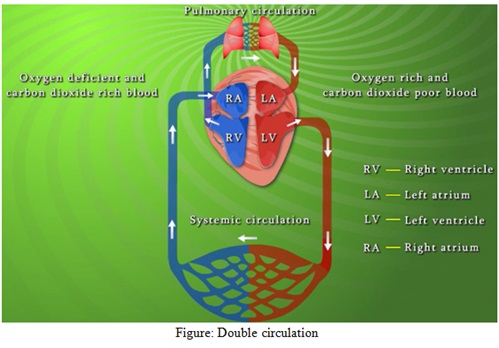
Q.6 What is meant by double circulation? What is its significance?
Solution: Double circulation refers to the process wherein blood passes two times through the heart in one complete cycle. In this circulation, the blood circulates in two distinct and separate pathways/loops (pulmonary circulation and systemic circulation). It is observed in birds and mammals as they have a four-chambered heart.
Pulmonary circulation: The pumping of blood from the heart to the lungs forms the pulmonary circuit. The right atrium of the heart receives deoxygenated blood from all the body parts. The deoxygenated blood then moves to the right ventricle. This deoxygenated blood is carried to the lungs from the right ventricle of the heart via the pulmonary artery. The blood is oxygenated in the lungs and is transported back to the left atrium by the pulmonary vein.
Systemic circulation: Flow of oxygenated blood, from the left ventricle of the heart, via arteries, arterioles and capillaries to the tissues is known as a systemic circuit. In the lungs, deoxygenated blood acquires oxygen and goes into the left atrium of the heart. From the left atrium, the oxygenated blood goes to left ventricle. Thereafter, the blood moves into the aorta and is carried to different body tissues by a network of arterioles, arteries, and capillaries. The deoxygenated blood is then collected from body tissues by a system of venules, veins and vena cava and emptied into the right atrium. This forms the systemic circulation. Systemic circulation provides nutrients, oxygen and other necessary substances to various body parts and carries back carbon dioxide and other harmful waste products away for elimination from the body.
Significance: The importance of double circulation is that it does not allow the mixing of oxygenated and deoxygenated blood. Thus, it ensures the efficient oxygen supply to the body organs.
Q.7 Write the differences between:
- (a) Blood and Lymph
- (b) Open and Closed system of circulation
- (c) Systole and Diastole
- (d) P-wave and T-wave
Solution:(a) Blood and Lymph
| Blood | Lymph |
| Blood is red due to the presence of haemoglobin in red blood cells (RBCs). | Lymph is colourless due to the absence of red blood cells (RBCs). |
| Blood contains RBCs, WBCs and platelets. | Lymph contains less number of WBCs and platelets. |
| Blood contains proteins. | It lacks proteins. |
| It transports digested food materials, respiratory gases and metabolic waste products. | It transports digested fats and fat-soluble vitamins. and is involved in the defence mechanism. |
| The flow of blood in the vessels is fast. | Lymph flows slowly. |
(b) Open and Closed system of circulation
| Closed system | Open system |
| In the closed type of circulatory system, blood flows inside the blood vessels all the time. | In the open type of circulatory system, blood is directly pumped into the body cavity called sinuses. |
| There are valves to prevent backward flow of blood. | There are no valves to prevent backward flow of blood. |
| Oxygen and nutrients diffuse out from the blood vessels to nourish the surrounding tissues/organs. | Oxygen and nutrients are directly supplied to tissues/organs by the blood present in the body cavity. |
| Blood and interstitial fluids do not mix. | There is no distinction between blood and interstitial fluid. They are called as hemolymph. |
| Blood flows at relatively higher speed. | Blood (hemolymph) flows at very slow speed. |
| Closed system is present in annelids and chordates. | Open type circulatory system is present in molluscs and arthropods. |
(c) Systole and Diastole
| Systole | Diastole |
| Systole refers to the contraction of heart muscles. | Diastole refers to the relaxation of heart muscles. |
| During systole, blood is pumped into the aorta and pulmonary arteries. Right ventricle contracts to send blood to the lungs via pulmonary artery. Left ventricle pumps blood into aorta. | During diastole, the heart chambers come back to original size to receive the blood. Blood received by atria is released into ventricles. |
| Atrio-ventricular valves close and semilunar valves open. | Atrio-ventricular valves open and semilunar valves close to prevent backflow of blood into the atria. |
(d) P-wave and T-wave
| P-wave | T-wave |
|
It indicates activation of Sino-atrial node.
|
It indicates ventricular relaxation.
|
| P-wave represents atrial excitation (depolarization) that results in atrial contraction. | T-wave shows the return of ventricles from excited to normal state (repolarization). |
Q.8 Describe the evolutionary change in the pattern of heart among the vertebrates.
Solution: The heart is a hollow muscular organ that pumps blood to all the tissues of the body. This process provides oxygen to various body parts. Evolution of heart has helped in more efficient transport of oxygen in the body by preventing the mixing of oxygenated and deoxygenated blood as described below:
- Fish: Fish have a very simple heart structure. Its heart is a two-chambered hollow tube-like structure. It has one atrium and one ventricle. Deoxygenated blood enters the atrium of the heart from where the blood is pumped into the ventricle. It then enters into the gills for oxygenation. The blood oxygenated in the gills is circulated across the body and finally, the deoxygenated blood from different body parts is transported back into the atrium of the heart.
- Amphibians: Amphibians have a three-chambered heart: two auricles and one ventricle. Oxygenated blood from the lungs enters into the left auricle while deoxygenated blood from other body parts enters into the right auricle. Both auricles transfer blood to the ventricle. In the ventricles, the oxygenated and deoxygenated blood mixes and this mixed blood is circulated to different body parts.
- Reptiles: Reptilians (except crocodiles) have an incomplete four-chambered heart. They have two auricles and a ventricle which is partially divided into two chambers. There is partial mixing of oxygenated and deoxygenated blood due to incomplete partitioning of the ventricles.
- Mammals and birds: Mammals and birds have a four-chambered heart. This prevents the mixing of oxygenated and deoxygenated blood. Right atrium receives deoxygenated blood and transfers it into right ventricle. From the right ventricle, deoxygenated blood is pumped into lungs to get oxygenated. Oxygenated blood is returned to left auricle of the heart. From left auricle, the oxygenated blood flows into the left ventricle and is pumped to whole body.
Q.9 Why do we call our heart myogenic?
Solution: Our heart is called myogenic heart because it receives signals for contraction from specialized cells called cardiac myocytes found in it. Cardiac myocytes form a specialized structure, known as nodal tissue, that possesses both muscular and nervous characteristics. The human heart receives the signals for contraction from the nodal tissue.
Q.10 Sino-atrial node is called the pacemaker of our heart. Why?
Solution: Sino-atrial node is called the pacemaker of heart because it is a specialized structure made up of cardiac myocytes that have both muscular and nervous characteristics. It is located in the upper side of right atrium. This structure has the ability to generate action potentials without any external stimuli. It initiates the impulse of contraction that subsequently spreads throughout the heart. It can generate a maximum of 70-75 action potentials per minute. It initiates and maintains rhythmic contraction of heart. Our heart usually beats around 70-75 times in a minute.
Q.11 What is the significance of atrioventricular node and atrioventricular bundle in the functioning of heart?
Solution: Atrio-ventricular (AV) node is present in right auricle at the base of inter-auricular septum, which separates right auricle from the ventricle. From this node, a bundle of nodal fibres called AV bundle arises and it passes via the AV septa. Immediately, it divides into left and right bundle. These bundles give rise to Purkinje fibres, which penetrate into the myocardium. These fibres along with the bundles form the structure called the bundle of His. This nodal system is auto-excitable, that is, it possesses the ability to generate an action potential in the absence of electric impulse. The electrical impulse is then passed on to ventricle by the bundle of His and Purkinje fibres. This impulse leads to contraction of the ventricles.
Q.12 Define a cardiac cycle and the cardiac output.
Solution: A complete cycle refers to the complete cycle of events from the start of one heartbeat to the start of next. It includes diastole and systole that involves filling of the heart with blood and then pumping it out. During diastole, the heart relaxes and fills with the blood. During systole, the ventricle contracts and pumps out the blood.
Cardiac output is the amount of blood pumped out by each ventricle in a minute. It is calculated by multiplying stroke volume (volume of blood pumped by each ventricle in a cardiac cycle) and heart rate (beats per minute).
Q.13 Explain heart sounds.
Solution: When the valves of heart open and close during the cardiac cycle, they produce a sound known as heart sound. These sounds are lub and dub. Lub is the first sound and is produced by the vibrations of the heart when bicuspid and tricuspid valves (valves present between atria and ventricle) close at the beginning of systole (when ventricular muscles contract). It is followed by second sound dub which is produced by vibrations caused due to closing of semilunar valves (which guard the pulmonary artery and the aorta) at the end of the systole.
Q.14 Draw a standard ECG and explain the different segments in it.
Solution: Electrocardiogram (ECG) is the graphical representation of the cardiac cycle. It is produced by an electrocardiograph. The normal ECG indicating that the heart is functioning properly is shown below.
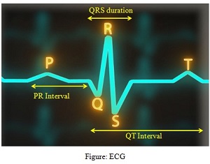
A typical human ECG includes the following waves:
• P-wave: It represents depolarization of the right and left atria. The impulse of contraction by the SA node is generated during this wave.
• QRS wave: It shows depolarization and contraction of the right and left ventricles. The contraction starts shortly after Q and indicates the beginning of systole.
• T-wave: It represents repolarization (return from excited to normal) and relaxation of the ventricles. Its end indicates the end of systole.
Waves P, R and T are above the base line and are called positive waves, while Q and S are below the base line and are known as negative waves.
More Resources of NCERT Solutions for Class 11 Biology
- NCERT Solutions for Class 11 Biology Chapter 1
- NCERT Solutions for Class 11 Biology Chapter 2
- NCERT Solutions for Class 11 Biology Chapter 3
- NCERT Solutions for Class 11 Biology Chapter 4
- NCERT Solutions for Class 11 Biology Chapter 5
- NCERT Solutions for Class 11 Biology Chapter 6
- NCERT Solutions for Class 11 Biology Chapter 7
- NCERT Solutions for Class 11 Biology Chapter 8
- NCERT Solutions for Class 11 Biology Chapter 9
- NCERT Solutions for Class 11 Biology Chapter 10
- NCERT Solutions for Class 11 Biology Chapter 11
- NCERT Solutions for Class 11 Biology Chapter 12
- NCERT Solutions for Class 11 Biology Chapter 13
- NCERT Solutions for Class 11 Biology Chapter 14
- NCERT Solutions for Class 11 Biology Chapter 16
- NCERT Solutions for Class 11 Biology Chapter 17
- NCERT Solutions for Class 11 Biology Chapter 18
- NCERT Solutions for Class 11 Biology Chapter 19
NCERT Solutions for Class 11 Biology Chapter 15 – FAQs
1. What is the difference between blood plasma and serum?
Plasma is the fluid portion of blood that contains clotting factors (like fibrinogen), while serum is the fluid that remains after blood has clotted - essentially plasma without the clotting factors. Plasma is obtained by centrifuging blood with anticoagulants, whereas serum is obtained after allowing blood to clot naturally.
2. How does the human heart prevent the mixing of oxygenated and deoxygenated blood?
The human heart has four chambers - two atria and two ventricles - that are completely separated by septa (walls). The right side handles deoxygenated blood while the left side handles oxygenated blood. The interventricular septum prevents mixing between the ventricles, and the valves (tricuspid, bicuspid, and semilunar) ensure unidirectional blood flow, maintaining complete separation of the two types of blood.
3. What is the difference between open and closed circulatory systems?
In an open circulatory system (found in arthropods and most molluscs), blood flows through open spaces called sinuses and is not always contained in blood vessels. In a closed circulatory system (found in annelids and vertebrates including humans), blood flows entirely through a network of blood vessels (arteries, veins, and capillaries) and doesn't directly bathe the tissues, with exchange occurring through capillary walls.
Q.1 Define growth, differentiation, development, dedifferentiation, redifferentiation, determinate growth, meristem and growth rate.
Ans-
Growth- The irreversible permanent increase in the size of an organ or its parts or even of an individual cell brought about by metabolic processes utilizing energy and nutrients over a period of time is called growth.
Differentiation- The process of maturation in which a cell converts into a highly specialized cell by a series of modification in the cell wall and cytoplasm to perform a particular function is called differentiation. For example, to form tracheal cells the cambial cells have to modify their cell wall and also loosen their protoplasm.
Development- The sequential and highly coordinated changes that an organism undergoes throughout their life cycle is called development. For example, in plants development means the changes which take place from seed germination to senescence.
Dedifferentiation- The phenomenon by which a differentiated cell performing specific function regains the capacity of division is termed as dedifferentiation. For example, the formation of meristems – interfascicular cambium and cork cambium from fully differentiated parenchymal cells. It happens only under certain conditions.
Redifferentiation- The process by which dedifferentiated tissues lose the capacity to divide again but mature to perform specific functions is called redifferentiation.
Determinate growth- When the growth of cell/ tissue/organ ceases after a specific size or dimension is attained it is called determinate growth or limited growth.
Meristem- The region in the plant body where actively dividing cells are present is called meristem. They accelerate the growth of plants and these tissues are named according to their location. For example, meristem at the shoot tip is called apical meristem, at the root tip is called root meristem and that in the stem is called lateral meristem.
Growth rate- The increased growth per unit time is termed as growth rate. The growth rate is expressed mathematically in terms of increase in size or number of cell per unit time. If the growth occurs as an increase in total surface area of a cell/ tissue without an increase in the number of cells it is called geometric growth. But when growth is accompanied by an increase in the total number of cells, it is called arithmetic growth.
Q.2 Why is not any one parameter good enough to demonstrate growth throughout the life of a flowering plant?
In plants, growth is the symbol of an increase in the quantity of protoplasm. Therefore, measuring the growth of protoplasm can demonstrate the growth of a plant. Like other organisms, plants also grow in various phases of their life cycle. The parameters to measure the growth of protoplasm vary for different parts of the plant; such as the parameters to measure the growth of fruit and seed are very different from each other. Some of the parameters are increased in the height, weight, length, diameter, surface area, volume, and cell number. Measuring growth involves the measurement of the increase of protoplasm in all these parameters. Thus, it is difficult to demonstrate growth throughout the life of a flowering plant using only one parameter.
Q.3 Describe briefly:
(a) Arithmetic growth
(b) Geometric growth
(c) Sigmoid growth curve
(d) Absolute and relative growth rates
Arithmetic growth: When a linear curve is obtained by plotting the growth parameter (for example root length) against time, it is called arithmetic growth. A typical example of arithmetic growth is elongation of root at constant rate. Arithmetic growth is the outcome of mitotic division, where one of the two daughter cells continue to divide while other differentiate and mature to perform specific function.
Mathematically, it is expressed as
Lt = L0 + rt
Where, Lt = length at the end of experiment
L0 = Length at the start of experiment
t = Time duration of experiment
r = Growth rate

Geometric growth: Geometric growth results from mitotic division where both the daughter cells retain the capacity to divide. Here, initially the growth is very slow which is called lag phase, but later it becomes very rapid which is called exponential phase. Later on, due to limited nutrient supply, cell division slows down again to attain stationary phase. If growth parameters are plotted against time for geometric growth, a sigmoid curve is obtained.

Sigmoid growth curve: During the early stages of development of plants (say at the time of seed germination), the growth rate is slow, but with time as the cell starts utilizing nutrient, the growth is very rapid and becomes exponential. Later on, when the number of cells increases and the amount of available nutrients become limited, the growth rate becomes stationary. When such growth is plotted against time an S-shaped curve called sigmoid curve is obtained.
Absolute and relative growth rates: Absolute growth rate is the measurement and the comparison of total growth per unit time. The relative growth rate is when the growth is expressed as an increase in specific parameter relative to its initial value per unit time known as relative growth rate.
Q.4 List five main groups of natural plant growth regulators. Write a note on discovery, physiological functions and agricultural/horticultural applications of any one of them.
Five main groups of natural plant growth regulators are:
- Auxin
- Gibberellin
- Cytokinins
- Ethylene
- Abscisic acid
Discovery of Auxin: The first observation of the presence of auxin in plants comes from the experiment of Charles Darwin and Francis Darwin on canary grass. They observed the bending of coleoptiles of canary grass towards the unilateral light source (phototropism). When the tip of the coleoptiles was cut, no bending was reported but when cut coleoptiles tip was placed over agar block it transmitted some chemical to the agar block. On placing this agar block over the cut coleoptiles, it again showed bending towards the unilateral source of light. Later on, in 1926, Auxin was isolated from the tip of coleoptiles of oat seedlings.
The physiological function of auxins: Shoot and root apices are the sites of auxin production in plants from where they are transported to their site of action. The auxins isolated from plants are indole-3-acetic acid (IAA) and indole butyric acid (IBA) while, NAA (naphthalene acetic acid) and 2, 4-D (2, 4-dichlorophenoxyacetic) are artificially synthesised auxins. The major physiological roles played by auxins are as follows:
- Promote apical dominance in plants
- Prevent premature falling of fruits and leaves
- Promotes abscission of older mature leaves and fruits
- Helps in xylem differentiation
- Promotes cell division
Agricultural/ horticultural application of auxins are:
- Used for root induction in cuttings when the plant is propagated through stem cutting
- Synthetic auxin 2,4-D is used as herbicides that selectively kills the dicotyledonous weeds without harming monocot plants
- Used for the development of seedless fruits as they promote parthenocarpy in tomatoes
- Sprayed on plants as they promote flowering for example in pineapples
Discovery of Gibberellins: Japanese farmers reported that few seedlings in rice field grow taller than others and never bears seeds; they called it “bakane” or foolish seedling disease. These seedlings were infected by a fungal pathogen, Gibberalla fujikuroi. Later on, E.Kurosawa, showed the reappearance of symptoms when the sterile filtrate was applied to uninfected plants and the active substance was later identified as gibberellic acid.
The physiological functions of Gibberellins:
- Help in breaking seed dormancy by activating the group of enzymes, hydrolyses, in the seeds which in turn utilises the stored nutrient.
- Determine the length of internodes
- Promote bolting in rosette leaves
- Delay senescence.
Agricultural/ horticultural application of Gibberellins:
- Delay fruit senescence, thus fruit remains on the tree for an extended period.
- Spraying Gibberellins increase the length of grapes stalks.
- In apple, it leads to elongation and improves the shape of fruits
- In Sugarcane crop, spraying of it increases the length of internode thus increasing yield.
Discovery of Cytokinins: Cytokinins were discovered by F. Skoog and his co-workers during tissue culture experiment of tobacco stem. They observed that the callus (undifferentiated mass of tissue) differentiate into plant only when it is supplemented with auxin along with coconut milk (or extract from vascular tissue, yeast extract or DNA). Skoog and Miller, latter were able to purify this substance, crystallized it and identified it as a cytokinesis promoting substance. They called it kinetin.
The physiological functions of Cytokinins-
- Synthesised in the region of rapid cell growth and promote cytokinesis
- Promote the formation of new leaves
- Enhance chloroplast formation in leaves
- Promote lateral shoot growth
- Delay leaf senescence by enhancing nutrient metabolism
Agricultural/ horticultural application of Cytokinins:
- Delaying senescence help in long-lasting flower which holds economic importance
- Differentiation of callus by application of cytokinins has great use in plant tissue culture, thus helps in cloning purpose
- Prevent apical dominance
Discovery of Ethylene: This is a gaseous hormone produced in large amount by ripening fruits. It was discovered by the observation that when ripen orange is kept with banana, it result in hastened ripening of bananas.
Physiological functions of ethylene:
- Shows the antagonistic effect of dormancy and break seed and bud dormancy
- Shows triple response in plants and stimulates shoot and root growth and differentiation
- Enhances leaf and fruit abscission
- Induction of femaleness in dioecious flowers
- Stimulates flower opening
- Enhances flower and leaf senescence
- Hastens fruit ripening
Agricultural/ horticultural application of Ethylene:
- Used to hasten fruit ripening
- Initiate flowering and synchronises fruit set in pineapples
- Used as an inducer of female flower in cucumbers thus increasing the fruit yield.
Q. 5 What do you understand by photoperiodism and vernalisation? Describe their significance.
Photoperiodism: The flowering in certain plants depends not only on the combination of light and dark exposures but also their relative duration. This response is called photoperiodism. It is the ability of the plant to detect and respond to the duration of light (length of day and night). Based on the flowering response of plants toward the length of light condition, they are divided into three classes:
Long Day Plants: These plants flower when the length of day (light duration) exceeds a critical duration, thus, in turn, they need a shorter dark period (night length).
Short Day Plants: For flowering, these plants need day length (light condition) less than a critical duration, thus, in turn, they need a long dark period (night) exceeding a critical duration.
Day-Neutral Plants: Plants in this group do not show any correlation between flowering and duration of light exposure.
Significance: Photoperiodism is a very important phenomenon in the life cycle of a plant as it affects the flowering of the plant. The plant does not flower if it does not receive certain day and light conditions and thereby is not able to complete the life-cycle. Understanding the phenomenon of photoperiodism is highly helpful in horticulture (flowering industry) for cultivating and obtaining flowers throughout the year. This is also an important feature in agriculture. Farmers choose the crop in a given area depending upon the photoperiodism response of the crop.
Vernalisation: The process of initiation of flowering or acquisition of a plants ability to flower in spring by exposure to prolonged cold or low-temperature conditions is called vernalisation. This ensures that reproductive development and seed production occurs in spring and summer, rather than in autumn. In such plants, low temperatures control the flower either in a quantitative or qualitative manner. Several cereal plants such as wheat, barley and rye have two varieties (spring and winter varieties) depending on their requirement for low temperature for flowering and grain filling. A similar phenomenon is observed in biennials herbs such as sugar beet and cabbage, which show vegetative growth in first season and flower and die in the second season following low-temperature exposure. In perennial plants, a period of cold is needed first to induce dormancy and then later, after a certain time frame, plants flower.
Significance: The process of vernalisation ensures that the plant has fully developed vegetative phase and is ready for flowering.
Q.6 Why is abscisic acid also known as stress hormone?
Abscisic acid (ABA) is one of the plant growth regulators which helps in increasing the tolerance of plants to withstand stress conditions such as:
- Water scarcity or high temperature: ABA stimulates the closure of stomata to control the water loss. This makes plant tolerant of such conditions.
- It favours seed dormancy and inhibits seed germination so that the seeds can withstand desiccation and other environmental conditions unfavourable for growth.
Thus, due to its role in stress tolerance ABA is called the stress hormone.
Q.7 ‘Both growth and differentiation in higher plants are open’. Comment.
Both growth and differentiation in higher plants are open. A plant continues to grow throughout its life by adding new shoot, branches, leaves, etc. Growth of a plant is brought about by the meristems located at different locations in the plant. The apical meristem results in the growth of root and shoot apices while lateral meristem increases the girth of the plant. The cells of these meristems have the capacity to divide and form specialized cells that make the plant’s body, while they also self-perpetuate. This type of growth where new cells are always being added to the plant body by the activity of the meristem is called open growth.
The cells derived from root and shoot meristems differentiate and mature to perform specific functions. This process is known as differentiation. In plants, differentiated cells undergo dedifferentitation under certain conditions wherein the cells which had lost the capacity to divide regain the capacity to divide again. During this process, meristems/tissues divide and produce cells that once again lose the capacity to divide but mature to perform a specific function. This process is known as redifferentiation. Thus the differentiation process is also open – cells/tissues arising out of the same meristem differ in the structure at maturity.
Q.8 ‘Both a short day plant and a long day plant can produce can flower simultaneously in a given place’. Explain.
The flowering in some plant takes place only when they get light exposure exceeding a critical photoperiod or dark period less than a critical duration; such plants are called long day plants. Similarly, some plants need the less day length or photoperiod and a long dark period exceeding certain critical duration are called short day plants. Both short day plant and long day plant can flower simultaneously in the same place if grown with adequate photoperiods by artificial means. For example, if both long day plant and short day plants are grown under long day condition (say during summer when day are longer) but the short day plants are shifted to dark after a critical photoperiod, then both long day plant and short day plant will flower simultaneously.

Q.9 Which one of the plant growth regulators would you use if you are asked to:
(a) induce rooting in a twig
(b) quickly ripen a fruit
(c) delay leaf senescence
(d) induce growth in axillary buds
(e) ‘bolt’ a rosette plant
(f) induce immediate stomatal closure in leaves.
(a) Auxin
(b) Ethylene
(c) Cytokinins
(d) Cytokinins
(e) Gibberellic acid
(f) Abscisic acid
Q.10 Would a defoliated plant respond to photoperiodic cycle? Why?
A plant where all the leaves are removed is called defoliated plant. A defoliated plant will not respond to photoperiodic cycle. This is because before flowering takes place, the shoot apices have to get modified into flowering apices. Flowering in plants depends on the specific duration of light and dark (photoperiod) which is perceived by leaves. According to the hypothesis, the hormonal substance necessary for flowering is synthesised in leaves in response to specific photoperiod and is transported to shoot apices to induce the formation of flowering apices. In a defoliated plant this hormonal substance is absent due to which they do not respond to photoperiodic cycle.
Q.11 What would be expected to happen if:
(a) GA3 is applied to rice seedlings
(b) dividing cells stop differentiating
(c) a rotten fruit gets mixed with unripe fruits
(d) you forget to add cytokinin to the culture medium.
Ans-
(a) Application of GA3 to rice seedlings results in an increase in the length between two nodes thus increasing the inter-nodal axis which makes the plant tall.
(b) If dividing cells stop differentiating, they form a callus. This may be due to the absence of cytokinin in the system.
(c) If a rotten fruit gets mixed with unripe fruits, it will produce ethylene and enhance the rate of ripening of unripe fruits.
(d) If we forget to add cytokinin to the culture medium, the cell will stop differentiating and form a callus.